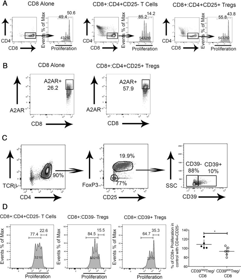
Fig. 7. Proliferation of hepatic CD8+ lymphocytes from Mdr2−/− mice is inhibited by Tregs in CD39-dependent manner.

CD8+ lymphocytes were labeled with the proliferation tracker Cell Trace Violet and incubated in the presence of IL-2 and plate-bound anti-CD3, and anti-CD28. CD8+ lymphocyte proliferation was evaluated by flow cytometry based determination of Cell Trace Violet dilution after incubation for 3 days. Generation 0 defining non-proliferating and Generations 1-4 the proliferating cells in the CD8 compartment are denoted in the histograms. CD8+ cells were cultured alone or in the presence of splenic CD4+CD25- Non-Treg lymphocytes or CD4+CD25+ Tregs at a ratio of 1:1 (A). Hepatic CD8+ cells from Mdr2−/− mice were cultured alone or in the presence of splenic CD4+CD25+ Tregs at a ratio of 1:1 for 3 days with subsequent evaluation of the adenosine receptor A2A expression on CD8+ cells (B). TCRβ+ CD4+ splenic lymphocytes were purified from adult Mdr2+/+ FoxP3-EGFP mice following treatment of i.p. IL-2c for 2 weeks and sorted by FACS into CD25-FoxP3- non-Tregs, CD39+, and CD39- Tregs (C) and co-cultured at a ratio of 1:1 with hepatic Cell Trace Violet labeled CD8+ T lymphocytes from adult Mdr2−/− mice for 3 days to determine percentage of proliferating CD8+ lymphocytes. CD8-proliferation in presence of CD39+ or CD39- Tregs was normalized against CD8-proliferation in co-cultures of CD8+ and non-Treg CD4+ cells assayed as internal standard in independent experiments. *p<0.05 in unpaired t test (D).
