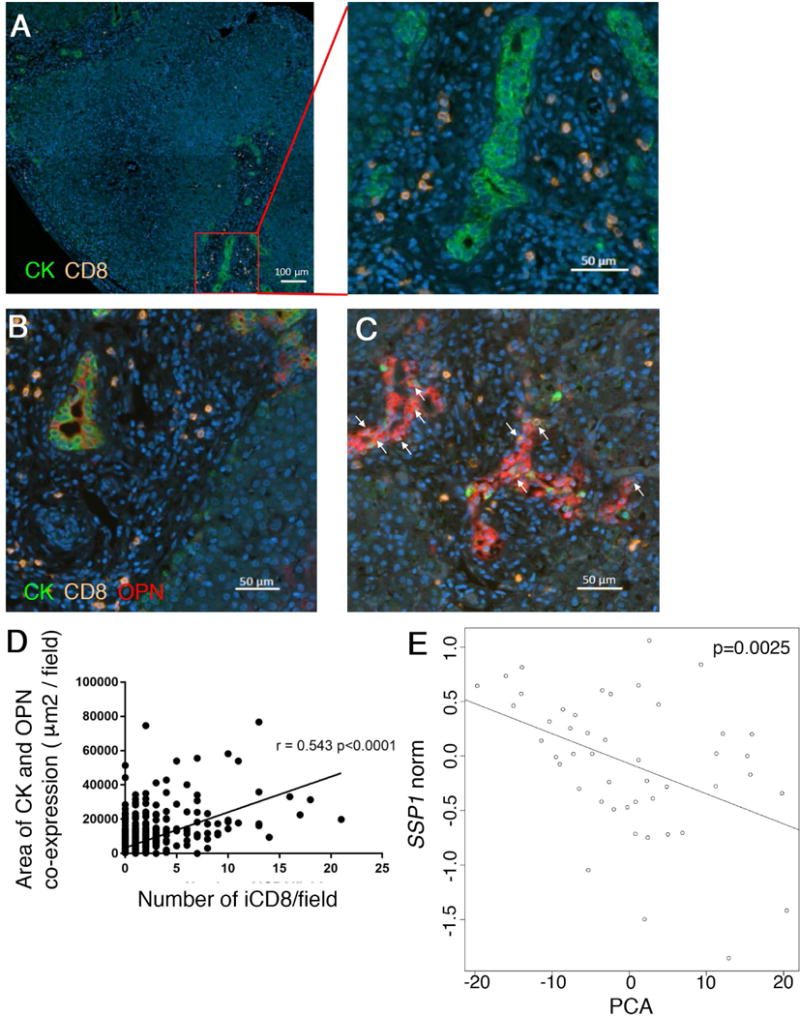
Fig. 8. Osteopontin expression by intrahepatic bile ducts is correlated with grade of infiltration by CD8 cells.

Archived FFPE liver sections from 8 patients with BA at diagnosis were subjected to multi-parameter immunofluorescence with antibodies against Pan-CK, CD8 and oteopontin. CD8 cells accumulate in the periportal area in BA, as shown by representative photomicrographs (A). CD8 cells infiltrating intrahepatic bile ducts (iCD8) are denoted by arrows and their number varies between patients and portal tracts. OPN expression by biliary epithelial cells is lower in samples without iCD8 (B) and up-regulated in presence of iCD8 (C). Co-expression of Pan-CK and OPN was plotted against the number of iCD8 per filed, as determined by image analysis. The p-value represents the Pearson’s correlation coefficient (D). Microarray gene expression data from a cohort of 47 patients with BA at diagnosis was analyzed for expression values for SSP1 (encoding OPN) and 120 Treg-associated genes. A negative correlation exists between SSP1 expression and a principal component analysis of Treg-gene expression values as predictors (E).
