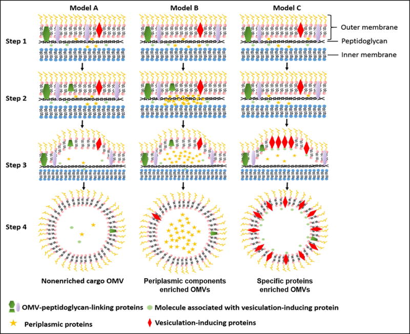
Figure 1. Biogenesis of OMVs.

Step1: Gram-negative bacteria cell envelop. In this stage, envelop proteins are homogenously distributed. Outer membrane is linked with peptidoglycan. Step2: Vesiculation initiation. The linking between outer membrane and peptidoglycan is lost through the movement of linking proteins or breaking the connection of outer membrane with peptidoglycan directly. Model A, B and C demonstrate three ways for OMVs production. Model A indicates the basal OMV production. Model B refers to the OMV production with enriched periplasma cargos. Model C shows the formation OMVs is located at specific proteins on the outer surface, and the dense proteins could induce the additional budding of OMV from gram-negative bacteria cell envelop.
