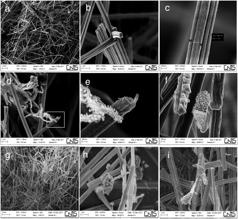
Figure 3.
Moonmilk deposit in the Etruscan Stanza degli Scudi, scanning electron microscopy (SEM). (a–c) Samples taken from the western wall (indicated with the letter d in Fig. 1a) in March 2017 were composed of needle fibre calcite and calcite nanofibres (moonmilk). (d–f) In the moonmilk samples from March, a microorganism, presumably Actinobacterium, was present; (e) is a magnification of picture (d). Note the nanoscale dimension of the mycelium. (g–i) Samples taken from the northern wall (indicated with the letter b in Fig. 1a) in December 2017. Of note, in (h), a microorganism was present similar to that identified in March on the western wall (compare (d), (e) and (h)). In (i), another microorganism, presumably an Actinobacterium strain, was present in the moonmilk.

