
Figure 5.
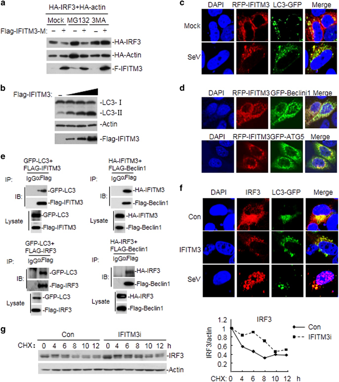
IFITM3 mediates autophagic degradation of IRF3. (a) Stable IFITM3-knockdown HEK293 cells were transfected with the indicated plasmids and treated with 3-MA or MG132 for 6 h before an immunoblot analysis was performed. (b) HEK293 cells were transfected with increasing amounts of Flag-IFITM3 and analyzed on immunoblots with the indicated antibodies. (c and d) HeLa cells transfected with the indicated plasmids were infected with SeV for 12 h or left untreated and then analyzed under immunofluorescence microscopy. (e) HEK293 cells were transfected with the indicated plasmids and applied to the coimmunoprecipitation and immunoblot experiments. (f) HeLa cells were transfected with Flag-IFITM3 and LC3-GFP plasmids, or infected with SeV. Immunofluorescence assays were performed using anti-IRF3 antibody as the primary antibody. (g) Stable IFITM3-knockdown HEK293 cells were infected with SeV and treated with CHX for the indicated times before immunoblot analysis was performed (left panel). The expression levels of IRF3 and actin were semi-quantified by measuring the grayscales of the bands on the western blots. The normalized expression of IRF3 is shown in the line chart (right panel).