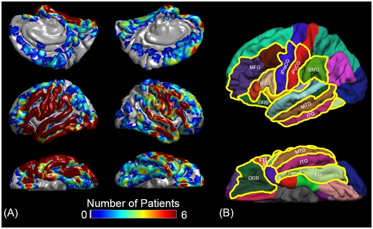
Figure 3.
Electrode coverage and regions of interest (ROIs). (A) The spatial extent of a total of 1,756 analyzed electrodes are indicated on the FreeSurfer’s average brain images17. (B) The boundaries of ROIs of the present study are denoted with yellow lines. MFG: middle-frontal gyrus. IFG: inferior-frontal gyrus (summation of pars opercularis [BA 44] and triangularis [BA 45]). ORB: orbitofrontal region (summation of pars orbitalis [BA 47] and lateral-orbitofrontal gyrus). iPreCG: inferior-precentral gyrus. iPoCG: inferior-postcentral gyrus. SMG: supramarginal gyrus. STG, MTG, and ITG: superior-, middle-, and inferior-temporal gyrus, respectively. FG: fusiform gyrus. The number of analyzed electrodes in each ROI was provided in Table 1.

