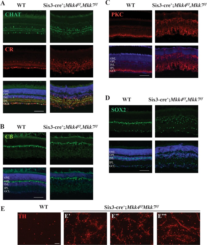
Fig. 7. Combined deficiency of Mkk4 and Mkk7 leads to severe alterations in retinal structure.
Representative sections of WT and Mkk4/Mkk7-deficient retinas stained with specific cell markers show clear disorganization of amacrine cells (a), slight defects in horizontal cells (b), and abnormal distribution and morphology of bipolar cells (c) and Müller glia (d) in retinas of Mkk4/Mkk7 double-deficient mutants. Merged images with DAPI (blue) are shown below. N ≥ 4 per genotype. e Mkk4/Mkk7 double-deficient flat mounts further display clumping and dendritic fasciculation of dopaminergic amacrine cells. N ≥ 4 per genotype. Scale bars: 100 μm

