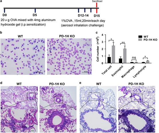
Figure 1.

PD-1H KO mice developed severe lung inflammation and mucus secretion in the OVA-induced asthma model. (a) Experimental protocol to induce experimental asthma: WT and PD-1H KO mice were immunized by i.p. injection of 20 μg OVA mixed with 4 mg aluminum hydroxide gel on days 0 and 5, followed by challenge with 1% OVA (15 ml/20 min/day) on three consecutive days (12–14) by aerosol instillation. The mice were killed for analyses on day 15. (b, c) BALF was collected and stained for leukocyte counts. (d) H&E staining of lung paraffin sections (e) PAS staining of lung paraffin sections for mucus-secreting goblet cells. The scale bars in b, d and e represent 100 μm. *P<0.05 and **P<0.01 (two-tailed student’s t-test). All values are expressed as means±s.e.m. N=8 per group. All experiments were repeated at least three times. BALF, bronchoalveolar lavage fluid; i.p., intraperitoneal; KO, knockout; OVA, ovalbumin.
