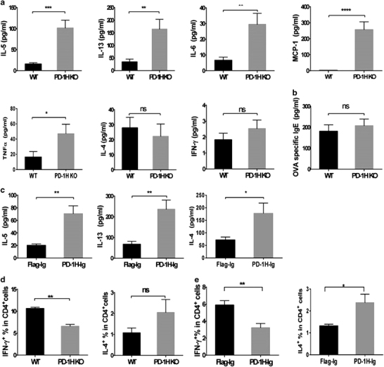
Figure 3.

Blockade of PD-1H enhanced innate and Th2-type cytokine production in OVA-induced asthma. (a) BALF from WT and PD-1H KO mice was collected, and the indicated cytokines were assessed by ELISA or CBA (N=8 per group). (b) Sera from the WT and KO mice was collected, and OVA-specific IgE was measured by ELISA (N=8 per group). (c) BALF from mice treated with the PD-1H-Ig or flag plasmid was collected, and the indicated cytokines were assessed by ELISA (N=8 per group). (d) Percentage of IL-4-producing and IFN-γ-producing CD4+ T cells in WT and KO mice in OVA-induced asthma. Lung lymphocytes isolated after the last OVA challenge were stimulated with PMA and ionomycin for 4 h in the presence of GolgiStop with brefeldin A. IL-4 and IFN-γ were measured by intracellular staining (N=8 per group). (e) The percentages of IL-4-producing and IFN-γ-producing CD4+T cells in wild-type mice treated with PD-1H-Ig or Flag-Ig plasmid in OVA-induced asthma were analyzed (N=8 per group). *P<0.05, **P<0.01, ***P<0.001 and ****P<0.0001 (two-tailed student’s t-test). All values are expressed as means±s.e.m. All experiments were repeated at least three times. BALF, bronchoalveolar lavage fluid; KO, knockout; OVA, ovalbumin; WT, wild-type.
