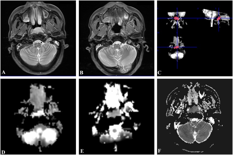
Figure 1.
A 48-year-old man with NPC in RG. Pre-treatment PdWI (A) showed the lesion located at the bilateral mucous membrane of the nasopharynx. No residual tumor was detected on axis PdWI after radiotherapy (B). 3D-ROI were manual drawing including the whole lesion on Kmean map (C). The Dmean (D) and Kmean(E) values were 1.36 × 10⁻³ mm2/s and 0.7 before treatment, respectively. The ADCmin (F) was 726.1 × 10⁻³ mm2/s before treatment.

