Figure 5.
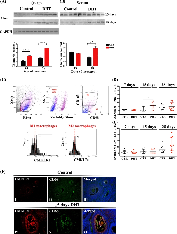
Chemerin and CMKLR1+ ovarian macrophages in DHT-treated rats. (A and B) Chemerin content in rat ovaries (A) and serum (B) after15 and 28 days of treatment with DHT. DHT-treated ovaries had higher chemerin content (P < 0.001 and P < 0.001, respectively) compared to control in both treatments (A). Serum chemerin (B) was significant higher in rats treated with DHT only after 28 days (P < 0.05), but not after 15 days. (C) Gating strategy used to identify ovarian M1 and M2 macrophages expressing CMKLR1. Side scatter (SS) and forward scatter (FS) were used to gate total ovarian leukocytes. M1 (CD68+CD163–) and M2 (CD68+CD163+ cells) macrophages were gated from viable leukocytes. The frequency of M1 and M2 macrophages expressing CMKLR1 was measured in comparison to the isotype control. M1 macrophages expressing CMKLR1 at 15 days of DHT treatment were significantly higher compared to control (P < 0.05), but not at 7 (P = 0.070) or 28 (P = 0.715) days (D). No differences were found in CMKLR1+ M2 macrophages in any day of DHT treatment [7 days (P < 0.713), 15 days (P < 0.715) 28 days (P < 0.057)] (E). (F) Double immunofluorescence using anti-CD68 (green; macrophages) and anti-CMKLR1 (red) on ovarian sections of 15 days DHT-treated rats. Low expression of CMKLR1 was observed in control ovaries (F, i–iii). The intensity of CMKLR1 staining was higher in the granulosa cells and CD68+ macrophages (arrows) from DHT-treated ovaries (F, iv-vi) compared to control. DAPI (nucleus; blue). Magnification of image (F): ×40.

