
Fig. 3:
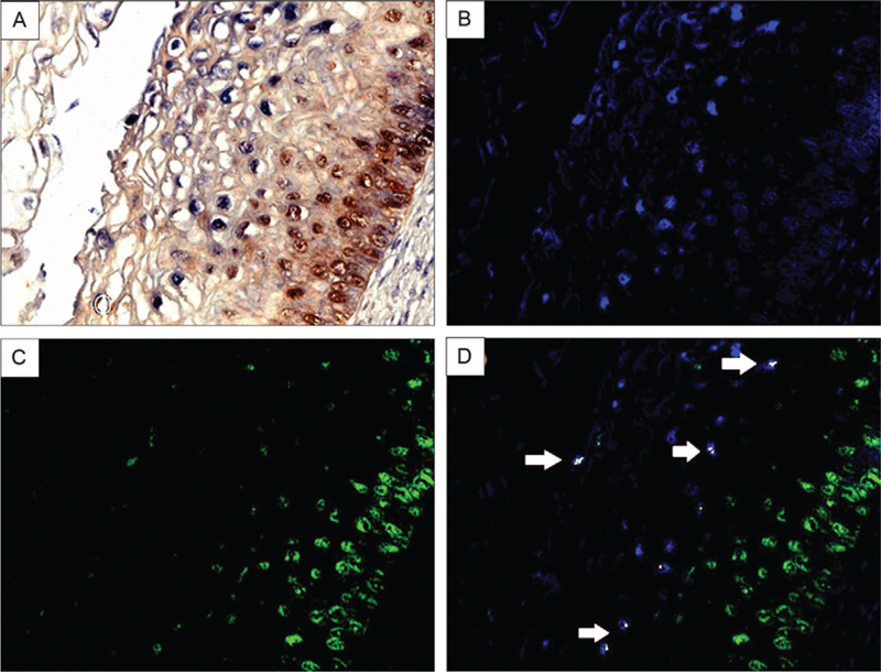
co-expression analysis of p16 INK4a and human papilloma virus (HPV) DNA 16 in low-grade cervical intraepithelial neoplasia (CIN). Co-expression analysis of HPV DNA 16 and p16INK4a in a CIN1 lesion. HPV DNA 16 was detected by in situ hybridization (blue signal), followed by co-expression analysis of p16 (brown signal) in the same tissue (A). The image was then analyzed by the computer-based NUANCE system which converted the HPV DNA signal to fluorescent blue (B) and p16 to fluorescent green (C). These latter two images are merged where cells with detectable HPV 16 and p16 would be fluorescent yellow (D); note the absence of any co-expression of these two targets where p16 dominates in the basal layer and HPV DNA is most abundant towards the cells at the surface of the CIN lesion.