Fig. 4.

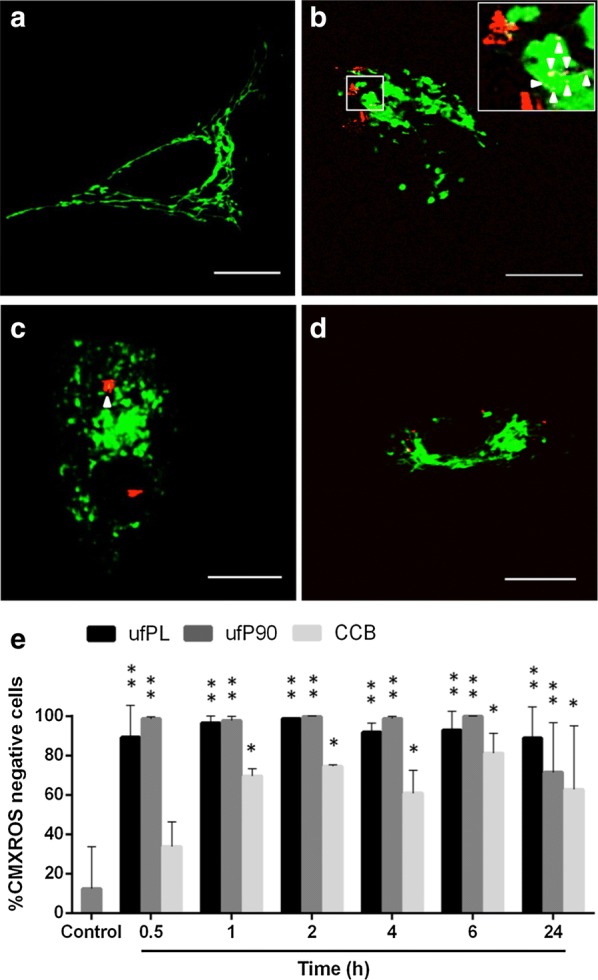
Mitochondrial damage by CBs in human lung fibroblasts. a–d Human lung fibroblasts (MRC-5 cell line) were exposed for 24 h to 20 µg/cm2 of three different types of CBs at 37 °C. Their mitochondrial organization was examined using CellLight® Mitochondria-GFP (green, Ex/Em 488/510 nm, ~ 3 µW radiant power at the samples) and CB particles were imaged under femtosecond pulsed illumination (red, 4 mW average laser power at the samples, emission detection: 400–410 nm in non-descanned mode). Co-localization between CBs and mitochondria is yellow due to the overlapping colors and additionally indicated by arrowheads. Representative images are shown from: a control condition (0 µg/cm2), scale bar: 15 µm; b incubation with ufPL particles, scale bar: 5 µm; c incubation with ufP90 particles, scale bar: 5 µm; d incubation with CCB particles, scale bar: 10 µm. e Time course study (0.5–24 h) of the loss of mitochondrial membrane potential after exposure to 20 µg/cm2 of three different types of CBs at 37 °C. After incubation, the cells were labeled with CMXROS fluorochromes and the percentage of CMXROS negative cells was determined. Data are represented as means ± SD (n = 3). Statistically different from control marked by *(p < 0.05) and **(p < 0.0005)
