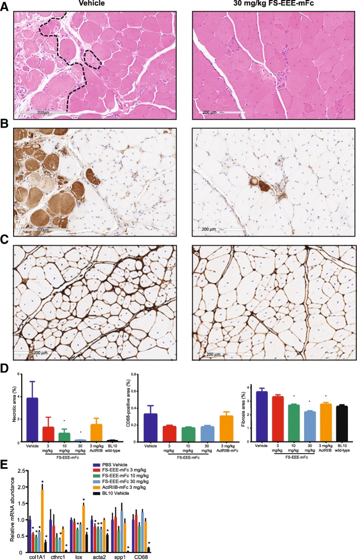
Fig. 3.
Histological staining and qPCR analysis of mdx quadriceps. a Representative images of hematoxylin and eosin staining depicting areas of heterogeneous necrosis from the vehicle control and 30 mg/kg FS-EEE-mFc. Dashed lines on the vehicle image depict manually defined boundaries of necrotic areas. b Representative images of mouse IgG-positive staining depicting areas of heterogeneous necrosis from the vehicle control (left) and 30 mg/kg FS-EEE-mFc (right). c Representative images of collagen I staining from vehicle control (left) and 30 mg/kg FS-EEE-mFc (right). d Total slide image analysis of IgG-positive staining for necrosis (left), CD68-positive staining for macrophage infiltration (center), and collagen I-positive staining for fibrosis (right). e qPCR of fibrosis and inflammation markers. *p < 0.05 compared to mdx vehicle-dosed group as described in the “Methods” section

