Fig. 5.
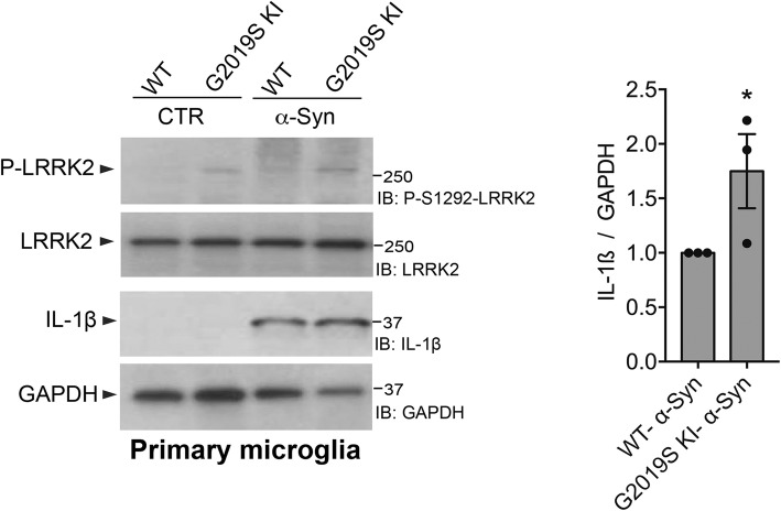
LRRK2G2019S KI primary microglia exhibit increased level of pro-inflammatory IL-1β after α-Syn pffs priming. LRRK2 WT and G2019S KI microglia lysates treated with 25 μM α-Syn pffs or PBS as control (CTR) were subjected to immunoblotting using P-LRRK2, total LRRK2, IL-1β, and GAPDH antibodies. Quantification of IL-1β is normalized for GAPDH. Data are representative of three independent experiments (bars represent the mean ± SEM; one-sample t test; *p < 0.05)

