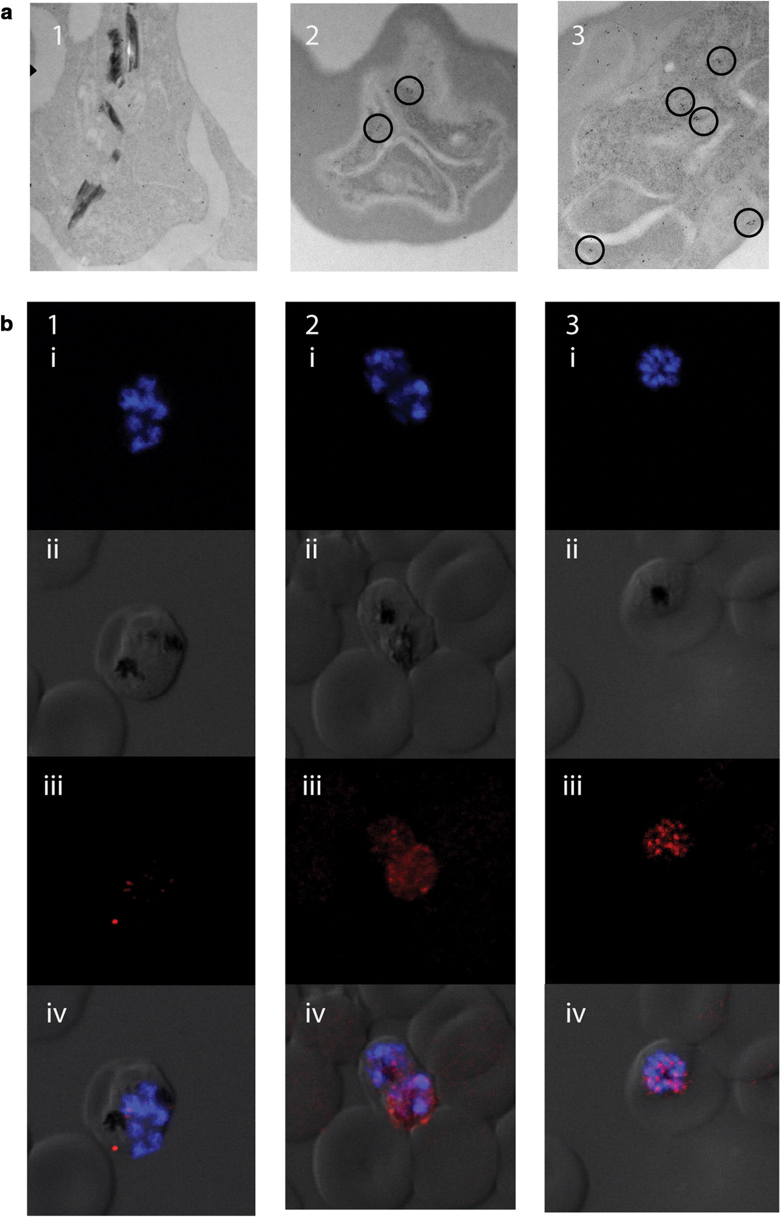
Fig. 2.

PfAtg18 forms puncta following DHA exposure a TEM images of Dd2R539T GFP-Atg18 parasites that were untreated (1) or exposed to 700 nM DHA for 10 min (2) or 1 h (3). Antibodies to GFP were gold-labelled (dots). Labels that congregate to form puncta are highlighted (circles). b Late-stage Dd2R539T Atg18-GFP parasites stained with DAPI (blue) and anti-GFP antibody (red). Rows depict images with (i) DAPI only, (ii) DIC only, (iii) anti-GFP only, and (iv) a merged image with all channels. Columns depict parasites that were untreated (1) or exposed to 700 nM DHA for 10 min (2) or 1 h (3)
