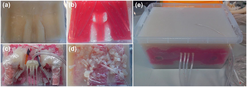
Figure 2:
(a–d) Photographs showing various stages during construction of the large anthropomorphic phantom, with: (a) an initial layer of fat TMM; (b) a layer of muscle TMM (red dye used to differentiate from other TMMs); (c) the prostate mimicking object in situ prior to surrounding TMM being deposited, as well as two ‘femur bones’ composed of bone TMM; (d) deposition of a heterogeneously distributed fat / muscle layer; and (e) the final phantom device. Dimensions: 280(w) × 390(l) × 200(h) mm3.

