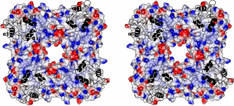
Fig.14.
Stereoview of surface diagram of the MDH tetramer with four fold axis passing through perpendicularly at the middle of the teramer. The positive, negative and neutral surface are colored as blue, red and white. The membrane binding segment is shown as a ribbon in black color. The three α-helixes are marked as α1,α2 and α3.

