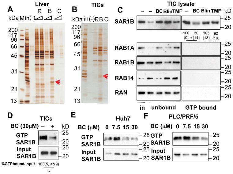
Figure 4. Baicalein blocks GTP binding of small GTPases in TICs.
Silver staining of proteins pulled down by R:Rosmarinic acid, B:Baicalin, C:Carnosic acid conjugated to nano-beads from protein lysates of A) liver and B) TICs. Blin-beads binding proteins are indicated by red arrows and are shown to include small GTPases by mass spectrometry. C) BC specifically blocks SAR1B from binding to GTP in TICs. TIC lysates were treated with BC, Blin and TMF at 30 μM for one hour in the presence of GTP-agarose. GTP bound proteins were pulled down and immunoblotted with indicated antibodies. SAR1B activation was quantitated by densitometry and expressed as percent of the control. Values are shown below each band. *p<0.05. Average (SEM) values from three experiments are shown. Statistical analyses were performed by One-way ANOVA with Bonferroni post-test. D) BC treatment in TICs blocked activation of SAR1B. TICs were treated with BC for 2 hr. The cells were lysed and GTP bound proteins were pulled down and immunoblotted with SAR1B antibody. SAR1B activation in TICs was quantitated by densitometry and expressed as percent of the control. Average (SEM) values from three experiments are shown. Statistical analysis was performed by the student’s t-test. *p<0.05. E) Huh7 and F) PLC/PRF/5 cells were treated with different concentrations of BC for 2 hr. The cells were lysed and GTP bound proteins were pulled down and immunoblotted with SAR1B antibody.

