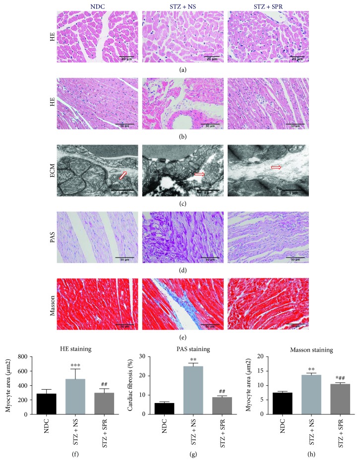
Figure 3.
SPR protected against myocardial hypertrophy and fibrosis in DCM. (a) Representative images of cardiac tissue stained with HE (transsection) for NDC, STZ + NS, and STZ + SPR groups (original magnification 40×). (b) Representative images of cardiac tissue stained with HE (longisection) for NDC, STZ + NS, and STZ + SPR groups (original magnification 20×). (c) Representative electron micrographs for the extracellular matrix from NDC, STZ + NS, and STZ + SPR groups. Compared to the NDC group, the deposition of the extracellular matrix (ECM) tended to be increased in the STZ + NS group, while SPR treatment reduced this pathological change. Blue arrows indicate ECM. (d, e) Representative images of cardiac tissue stained with PAS and Masson for the NDC, STZ + NS, and STZ + SPR groups are presented (original magnification 20×). (f, e) Quantification results summarizing the cross-sectional diameter of myocytes within transverse cardiac sections. (g, h) Quantification results of the PAS and Masson staining positive area in cardiac sections of rats treated under the conditions indicated, respectively. N = 8 for each group, data are shown as means ± SEM. ∗P < 0.05, ∗∗P < 0.01, and ∗∗∗P < 0.001 vs. NDC group; ##P < 0.01 vs. STZ + NS group.

