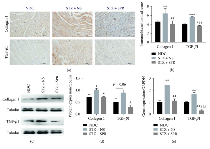
Figure 4.
SPR protected against myocardial hypertrophy and fibrosis in DCM. (a) Representative immunohistochemical micrographs of cardiac tissue stained with collagen 1 and TGF-β1 (original magnification 20×). (b) The IHC scores of heart sections for collagen 1 and TGF-β1 IHC staining. (c) The protein expression of collagen 1 and TGF-β1 examined by Western blot. (d) The quantification data of Western blot for collagen 1 and TGF-β1. (e) Gene expression of collagen 1 and TGF-β1 for these 3 groups. N = 8, data are shown as means ± SEM. ∗P < 0.05, ∗∗P < 0.01, and ∗∗∗P < 0.001 vs. NDC group; #P < 0.05, ##P < 0.01, and ###P < 0.001 vs. STZ + NS group.

