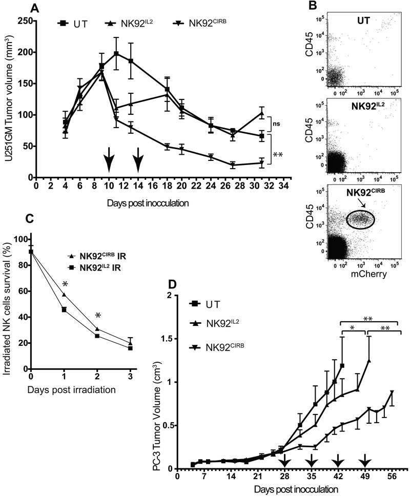
Figure 6.
NK92IL2 and NK92CIRB cells evaluation in vivo. A, When U251 tumor volume reached ~160mm3, non-irradiated NK92IL2 and NK92CIRB cells (107 cells in 200ul per mouse) were injected into mice (arrows), via the tail vein. A second injection of non-irradiated NK cells (5×10⁶ cells) was carried out 4 days later. Tumor sizes were monitored until 31 days post tumor implantation. B, 17 days after the last NK cells injection, animals were sacrificed and blood was collected from 3 animals in each group. Blood samples (0.5ml) were processed and analyzed by flow cytometry using human specific marker CD45 and the mCherry fluorescence marker, which is co-expressed with IL2 or CIRB in NK92IL2 and NK92CIRB, respectively. NK92CIRB cells detected (circled). C, Cell survival of NK cells irradiated in vitro at 10Gy (0.83gy for 12 min) and then plated in complete NK92 media was determined using Trypan Blue exclusion every 24 hours. The survival advantage of NK92CIRB cells was statistically significant at days 1 and 2 (One-way Anova test *P<0.05). D, anti-tumor efficacy of irradiated NK92IL2 and NK92CIRB cells in vivo against PC-3 tumors grown in 5 week-old male Nod/Scid mice. When tumor size reached ~200 mm3, NK cells were irradiated with 500cGy were administered as four weekly injections of 15×106 cells in 200ul per mouse, via the tail vein (arrows). After the last NK92CIRB cells injection, a significant tumor growth delay of 17 days was recorded (**P<0.01), comparatively to the untreated group. NK92IL2 treated group tumors produced a tumor delay of only 7 days (*P<0.05). Statistical differences were determined using One-way Anova test.

