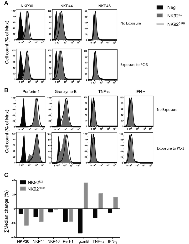
Figure 7.
NK92CIRB and NK92IL2 cells activation by direct contact with PC-3 cancer cells. NK cells and PC-3 cells were co-cultured for a duration of 3 hours after which their expression profile of A, NKP30, NKP44, NKP46 and B, Perforin-1, Granzyme-B, TNF-α and IFN-γ, were compared to NK cells grown alone. C, ∑Median expression of histograms by FlowJo software of the data in A, B and supplementary Table S2. Data show the reduction in surface expression for NKP30, NKP44, and NKP46. In NK92IL2 Perforin-1, Granzyme-B, TNF-α and IFN-γ decreased suggesting release while they increased in NK92CIRB (36%, 21% and 16%, respectively) implying replenishment of these effectors. After 3 hours of contact with cancer cells, NK92CIRB cells still retained a distinct advantage by harboring all receptors and effectors in excess over NK92IL2 (supplementary Table S2).

