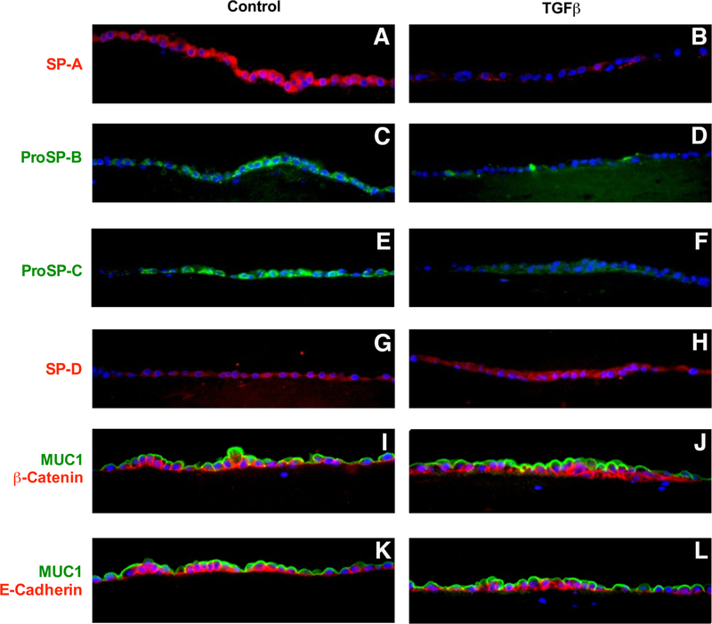
Figure 2. Immunocytochemistry of the effect of TGF beta on SP-A, proSP-B, proSP-C, and SP-D.
Type II cells were cultured under ALI conditions with and without TGF beta for last four days as in Figure 1. The exposures were set for the ALI control conditions, and the TGF beta condition was photographed at the same exposure and there was no computer enhancement of the images. Panel A SP-A, Panel B SP-A with TGF beta, Panel C proSP-B, Panel D proSP-B with TGF beta, Panel E proSP-C, Panel F proSP-C with TGF beta, Panel G SP-D, Panel H SP-D with TGF beta, Panel I Muc 1 (green) and beta catenin (red), Panel J Muc 1 (green) and beta catenin (red) with TGF beta, Panel K Muc 1 (green) and E-cadherin (red), Panel L Muc 1 (green) and E-cadherin (red) with TGF beta. These results are representative of four independent experiments

