
Figure 1 |. Filamentous tau pathology of Pick’s disease.
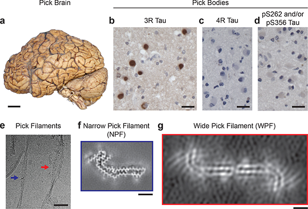
a, The brain used for cryo-EM (patient 4) showed atrophy of anterior frontal and temporal lobes of the cerebral cortex. Scale bar, 5 cm. b-d, Staining of Pick bodies in the frontotemporal cortex of patient 4 by antibody RD3 (3R tau; brown) (b), but not by antibodies Anti-4R (4R tau) (c) or 12E8 (pS262 tau and/or pS356 tau) (d). Nuclei were counterstained blue. Scale bars, 20 μm. e, Cryo-electron micrograph of tau filaments extracted from grey matter of the frontotemporal cortex of patient 4, in which narrow (NPFs; blue arrow) and wide (WPFs; red arrow) Pick filaments could be distinguished. Scale bar, 500 Å. f, Unsharpened cryo-EM density of NPF from patient 4. Scale bar, 25 Å. g, Unsharpened cryo- EM density of WPF from patient 4. Scale bar, 25 Å.