
Figure 1 |. Further characterization of the filamentous tau pathology of Pick’s disease.
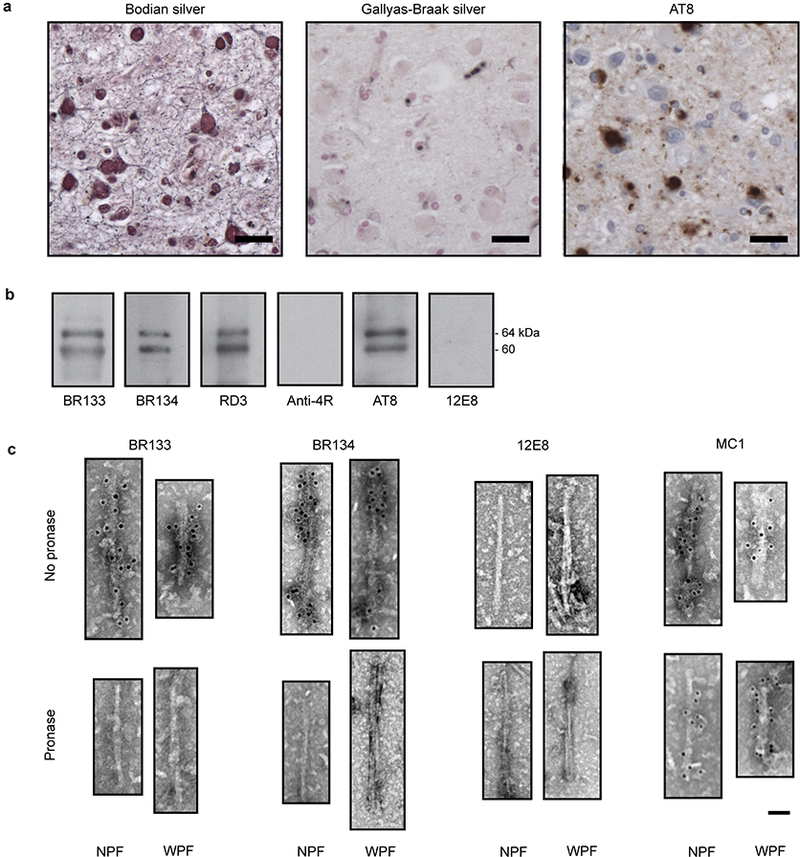
a, Staining of Pick bodies in the frontotemporal cortex of patient 4 by Bodian silver and antibody AT8 (pS202 and pT205 tau), but not by Gallyas-Braak silver. Nuclei are counterstained blue. Scale bars, 20 μm. b,c, Immunolabeling of the tau filaments extracted from the frontotemporal cortex of patient 4. Immunoblots (b) with antibodies BR133 (tau amino-terminus), BR134 (tau carboxy-terminus), RD3 (3R tau), Anti-4R (4R tau), AT8 (pS202 and pT205 tau) and 12E8 (pS262 tau and/or pS356 tau). Immunogold negative-stain electron micrographs (c) with antibodies BR133, BR134, 12E8 and MC1 of NPFs and WPFs with and without pronase treatment. Scale bar, 500 Å.