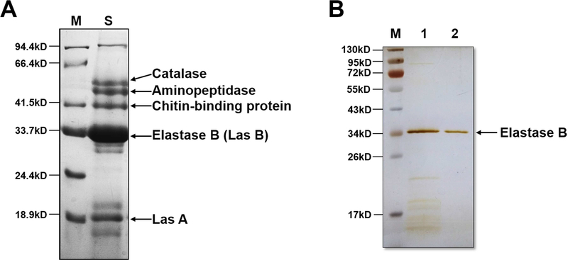
Figure 1. Identification of the PAO1 secreted proteins and purification of elastase B.
(A) The secreted proteins of PAO1 was separated by SDS-PAGE, visualized by Coomassie Brilliant Blue R-250, followed by in-gel trypsin digestion and mass spectrometry analysis. The identified proteins are indicated. Lane M, marker proteins; lane S, PAO1 secreted proteins. (B) Lane 1, fraction collected from 100 mM NaCl elution on the second run of Q-Fast Flow Sepharose column. Lane 2, purified elastase B from the HPLC gel filtration column. The protein bands were visualized by silver staining.

