Figure 5.
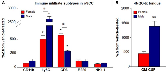
Increased immune cell infiltrate during the development of 4NQO-induced oSCC. (A) Using flow cytometry, infiltrating immune cell subpopulations were quantified in dissociated tongue tissue from female (red bars) and male (blue bars) vehicle-treated mice (N = 9 females, 9 males) and mice with 4NQO-induced oSCC (N = 8 female, 10 males). Data are presented as a percent change from vehicle-treated mice. The percent of total live cells quantified for both vehicle and 4NQO-treated mice with oral SCC are available in Table 2. P < 0.05 for Sex vs. Treatment by Two-way ANOVA; *P < 0.05, **P < 0.01 for Holm-Sidak post hoc comparisons for treatment; #P < 0.05 for Holm-Sidak post hoc comparisons for sex. (B) Granulocyte macrophage-colony stimulating factor (GM-CSF) protein was measured in homogenized tongue tissue from male (N = 5) and female (N = 4) mice with 4NQO-induced oSCC compared to vehicle-treated male (N = 4) and female (N = 4) mice. GM-CSF protein concentration was significantly higher in male mice (blue bar) with 4NQO-induced oSCC compared to female mice (red bar). Data are represented as percent change from vehicle-treated mice. **P < 0.01 by Unpaired Student’s t-test.

