Figure 2.
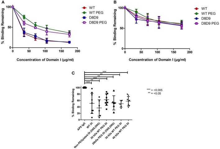
PEGylated DI blocks binding of IgG in APS serum samples to whole β2GPI. (A) This graph shows the combined results from inhibition ELISAs of serum samples from three patients whose binding to whole β2GPI was inhibited more strongly by non-PEGylated than by PEGylated DI constructs. (A) Contains patients with a high level of serum aDI antibodies (average >80 GDIU). (B) This graph shows the combined results from inhibition ELISAs of serum samples from three patients whose binding to whole β2GPI was inhibited equally by non-PEGylated and PEGylated DI constructs. These patients have a lower level of serum aDI (average < 45 GDIU). (C) The dot bot shows the combined results from testing samples from 9 patients [including all six from (A,B)]. The first column on the left shows binding to β2GPI in the absence of any inhibitor and the other columns show the binding in the presence of different inhibitors at a concentration of 100 μg/ml. For both WT-DI and DI(D8S,D9G), addition of 20 kDa PEG does not alter the inhibitory capacity of DI in this assay and the same is true for 30 and 40 kDa PEG in the case of WT-DI (these larger PEG sizes were not tested for DI(D8S,D9G). Significant differences were seen between the results obtained with APS IgG alone and those obtained with all inhibitors PEGylated or non-PEGylated (***p < 0.005, **p < 0.05) but no significant differences were seen between any of the inhibitors.

