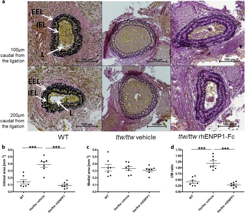
Fig. 4. Administration of rhENPP1-Fc prevents intimal proliferation after carotid ligation in ttw/ttw mice.
RhENPP1-Fc treatment was started 7 days prior to carotid ligation, and serial sections of the left carotid arteries were taken 14 days after carotid ligation. Histological analysis (Von Gieson’s stain) was performed on sections taken either 100 (upper panel) or 200 (lower panel) µm from the point of ligation from WT, vehicle-treated ttw/ttw or rhENPP1-treated ttw/ttw- mice, shown from left to right (a). The internal elastic lamina (IEL), external elastic lamina (EEL), and lumen (L) are indicated by arrows. The scale bar represents 100 µm. Morphometric quantitation was performed on intimal (b) and medial (c) areas, and the I/M ratio was calculated (d). Values are presented as the mean ± SEM, n = 7 each group, ***p < 0.001 (one-way ANOVA multiple group comparison followed by the Bonferroni’s post hoc test)

