Figure 4.

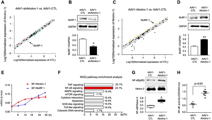
Atrolnc‐1 upregulates MuRF‐1 expression in C2C12 myotubes. (A) Scatter analysis of Myogenesis & Myopathy PCR Array identified that MuRF‐1(arrowhead) was downregulated in C2C12 myotubes when Atrolnc‐1 was knocked down by AAV1‐shAtrolnc‐1. AAV1‐carried scrambled shRNA (AAV1‐CTL) served as control. Dash lines indicate 2‐fold changes. (B) The down‐regulation of MuRF‐1 was confirmed by immunoblots in C2C12 myotubes with Atrolnc‐1 knockdown, GAPDH was used as loading control (mean ± SEM; n = 3, *P < 0.05). (C) Scattered plot of Myogenesis & Myopathy RT‐PCR Array in C2C12 myotubes with Atrolnc‐1 overexpression (AAV1‐Atrolnc‐1), MuRF‐1 was indicated with the arrowhead. AAV1 carried scrambled shRNA (AAV1‐CTL) served as control. Dash lines indicate 2‐fold changes. (D) Immunoblots confirmed the up‐regulation of MuRF‐1 in C2C12 myotubes with Atrolnc‐1 overexpression (mean ± SEM; n = 3, **P < 0.01). (E) The time course of Atrolnc‐1 and MuRF‐1 expression in response to serum depletion. The mRNA level was examined using RT‐PCR. β‐actin was used as internal control. SF, serum free (mean ± SEM; n = 3 in each group). (F) KEGG pathway enrichment analysis of genes altered by the knockdown or overexpression of Atrolnc‐1. NF‐κB signalling (red) was linked to the Atrolnc‐1 expression. (G) Immunoblot shows NF‐κB (p65) was increased in the nuclear fraction of C2C12 myotubes with Atrolnc‐1 overexpression (mean ± SEM; n = 3/group, *P < 0.05). (H) NF‐κB activity assay revealed overexpression of Atrolnc‐1 activated NF‐κB signalling in C2C12 myotubes (mean ± SEM; n = 8 in each group).
