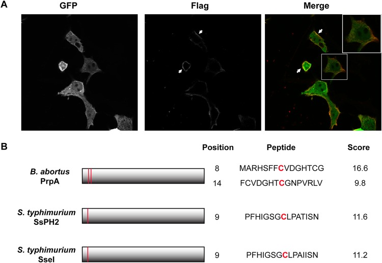
FIG 1.
PrpA is translocated to the cell membrane. (A) HEK293 cells were transfected with pEGFP-PrpA3×Flag and examined by indirect immunofluorescence confocal microscopy using an anti-Flag monoclonal antibody under nonpermeabilization conditions. Green, GFP; red, Flag. (B) Schematic representation of B. abortus PrpA and S. enterica serovar Typhimurium SseI and SspH2, highlighting the predicted palmitoylation motifs and their scores.

