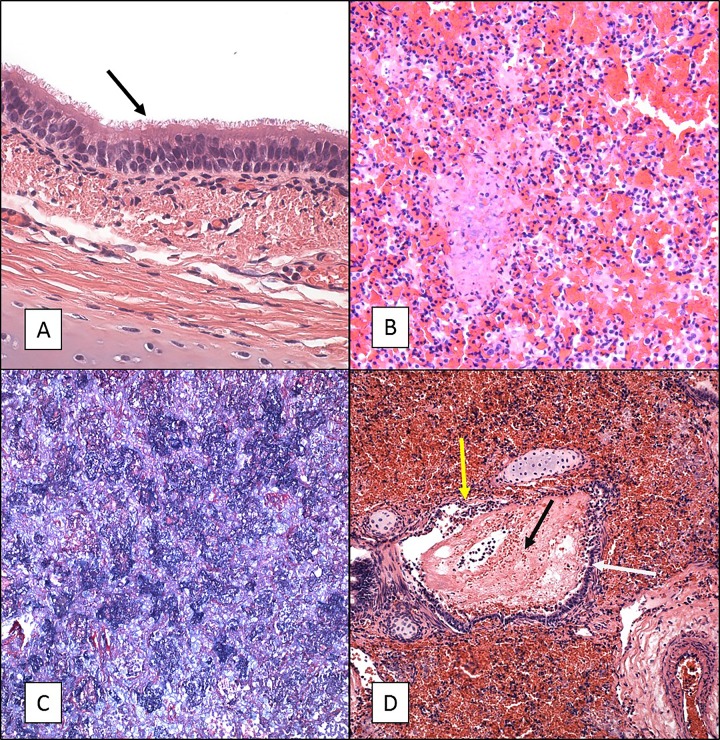
FIG 1.
H&E and PTAH staining of tissue sections from 5- to 6-week-old baboons infected with B. pertussis. Five- to 6-week-old baboons were challenged with B. pertussis. H&E- and PTAH-stained slides were prepared from tissues as described in Materials and Methods. Representative images are presented. (A) Tracheal tissue stained with H&E showing intact cilia (arrow) and a normal appearance. (B) Lung tissue stained with H&E showing severe acute vascular leakage and mostly acute inflammation. (C) PTAH staining of lung tissue indicating intra-alveolar fibrin deposition in blue. (D) H&E staining of lung tissue showing necrotizing bronchitis/bronchiolitis. White arrow, bronchus, intact mucosa; yellow arrow, damaged mucosa; black arrow, intralumenal blood and edematous fluid. Magnifications, ×600 (A) and ×200 (B to D).

