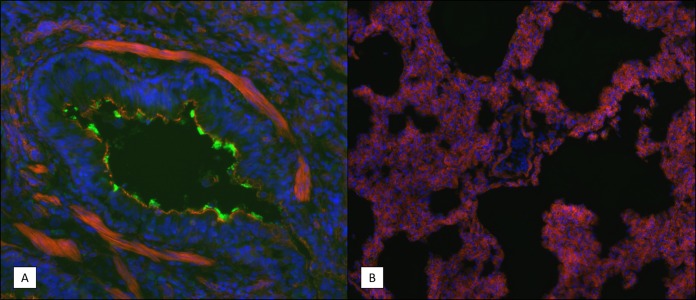
FIG 9.
Immunohistochemistry (IHC) staining of B. pertussis in lung tissue sections. Frozen lung tissue sections were prepared from infected animals on day 7 postchallenge as described in Materials and Methods. B. pertussis bacteria were visualized using rabbit polyclonal antibodies specific for a surface-exposed antigen. Representative images are shown. Blue indicates the DAPI staining of the nucleus, red indicates the staining of F actin, and the green in panel A indicates B. pertussis bacterial cells. (A) Bronchiole showing bacteria lining the mucosal surface. (B) Alveolar space of an infected animal showing an absence of bacteria. Magnifications, ×200 (A) and ×100 (B).

