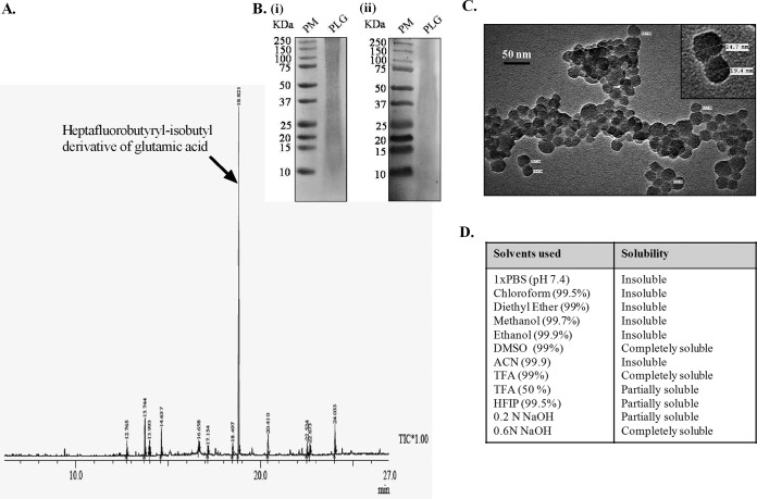
FIG 1.
Chemical and physical characterization of PLG isolated from M. tuberculosis cell wall. (A) GC-MS chromatogram of the PLG peptides. The arrow indicates the heptafluorobutyryl-isobutyl derivative of glutamic acid. Rt, retention time. (B) Tricine SDS-PAGE (i) and immunoblot analysis (ii) of PLG. For immunoblotting, primary antipolyglutamine monoclonal antibodies were employed. PM, Precision Plus protein standards. (C) TEM micrographs of purified PLG. The inset displays the same at higher magnification. (D) Table shows the solubility profile of M. tuberculosis PLG in various organic and inorganic solvents.

