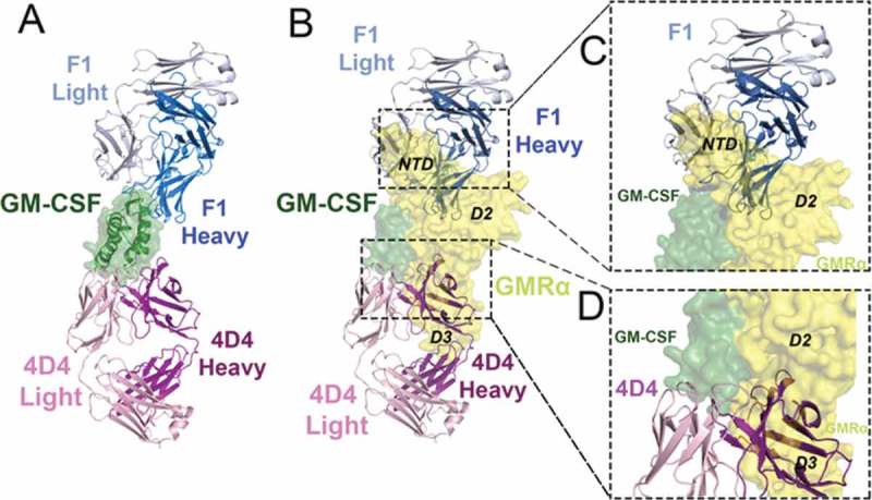
Figure 3.

The F1 and 4D4 Fabs bind to opposed surfaces of GM-CSF but both disrupt GM-CSF interaction with GMRα. (A) Cartoon representation of the GM-CSF:F1 Fab and GM-CSF:4D4 Fab structures superimposed via GM-CSF (shown as a cartoon with a transparent molecular surface overlay). Molecules are colored; F1 Fab heavy chain (blue), F1 light chain (grey), 4D4 heavy chain (purple), 4D4 light chain (light pink) and GM-CSF (green). (B) The F1 and 4D4 Fab complexes from (A) were superimposed, via GM-CSF, with the GM-CSF:GMRα binary complex structure (PDB ID: 4RS1).10 The GM-CSF:GMRα binary complex is shown as a molecular surface, with GMRα in yellow. (C) Close-up view of the steric clash between the F1 Fab heavy and light chain variable domains with the GMRα NTD and D2 in the GM-CSF:GMRα binary complex. (D) Close-up view of the steric clash between the 4D4 Fab heavy chain variable domains with GMRα D2 and D3 in the GM-CSF:GMRα binary complex.
