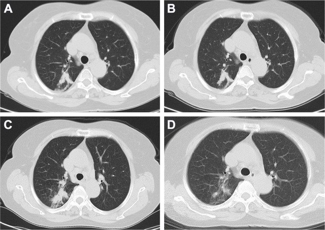
Figure 1.
CT image.
Notes: (A) Infiltration of the right upper lobe of the lung before treatment. (B) Infiltration of the right upper lobe of the lung after IL-2 therapy. (C) Infiltration of the right upper lobe of lung worsened after PD-1 inhibition. (D) Infiltration of the right upper lobe of lung absorbed after 6 weeks of anti-TB treatment.
Abbreviations: CT, computed tomography; TB, tuberculosis.

