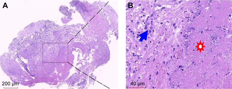
Figure 2.
Histopathological findings of TB granuloma from the lung biopsy.
Notes: A large amount of caseous necrosis surrounded with epithelioid cells and diffused infiltrating lymphocytes (paraffin-embedded tissue by H&E staining). (A) Original magnification (20×). (B) Local magnification of (A) (400×). Solar marking: caseous necrosis; blue arrows: epithelioid cells.
Abbreviation: TB, tuberculosis.

