Figure 6.
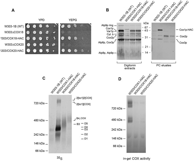
Cox18p and Cox20p are components of the Cox2p module. A, the parental WT, the cox18 and cox20 null mutants, and the null mutants expressing HAC tagged Cox18p and Cox20p were serially diluted and spotted on rich glucose (YPD) and rich ethanol plus glycerol (YEPG). The plates were incubated for 2 days at 30 °C. B, mitochondria of the WT and the stains expressing HAC-tagged Cox1p (MRSIO/COX1-HAC), Cox18p (W303/COX18-HAC), and Cox20p (W303/COX20-HAC) were grown in YPGal without additional growth in medium containing chloramphenicol. Mitochondria were prepared and labeled as in Fig. 3A. The digitonin extracts and eluates from the protein C antibody beads (PC eluates) were separated by SDS-PAGE on a 12% polyacrylamide gel. C, the fractions of B eluted from the PC beads were separated by BN-PAGE on a 4–13% polyacrylamide gel, transferred to a PVDF membrane and exposed to X-ray. D, same as C except that the gel was stained for COX activity.

