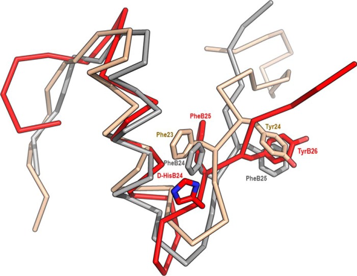
Figure 2.
An overlay of the B chains of human insulin with human IGF-1 and [d-HisB24]-insulin. Insulin (PDB code 1MSO, crystal structure) is shown in gray, IGF-1 (PDB code 1GZR, crystal structure) is ocher, and [d-HisB24]-insulin (PDB code 2M2P, NMR structure with the lowest energy at pH 8) is red. Positions of downshifted d-HisB24, PheB25, and TyrB26 in [d-HisB24]-insulin are shown together with corresponding residues in insulin (PheB24 and PheB25) and IGF-1 (Phe23 and Tyr24).

