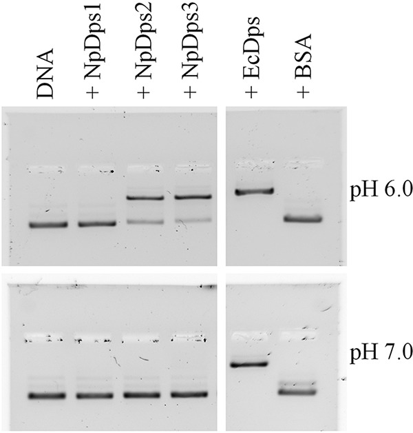
Figure 2.

EMSA to analyze the DNA-binding properties of the NpDps proteins under different pH conditions. At pH 6.0 and 7.0, 125 ng of plasmid DNA (pSB1A3 vector) was incubated with 1 μg of each Dps protein, and separated on an agarose gel (1%). E. coli Dps (EcDps) and BSA served as a positive and negative control, respectively. Plasmid DNA is shown in lane DNA. Thiazole orange staining was performed after gel electrophoresis for DNA detection. The gel documentations are shown in inverted colors.
