Figure 2.
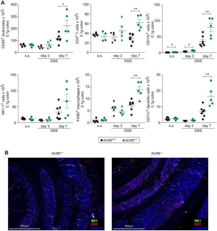
Enhanced immune cell infiltration into the colon of Nr2f6−/− diseased mice. (A) Colon-infiltrating cell numbers per 0.1 g of tissue of wild-type and Nr2f6−/− mice were stained for CD45+ leucocytes (p=0.029, day 7), CD3+ T cells (p=0.0008, day 7), CD11b+ cells (p=0.023, day 0; p=0.022, day 3; p=0.0072, day 7), NK1.1+ natural killer cells, F4/80+ macrophages (p=0.0028, day 7) and CD11c+ dendritic cells (p=0.0065, day 7) at indicated time points (n=5–7). (B) Representative immunofluorescence staining for CD3+ T cells (red), NK1.1+ natural killer cells (green) and Hoechst nuclear stain (blue) of wild-type and Nr2f6−/− Swiss rolls (n=5). Data are presented as mean±SEM error bars and are representative of at least two independent experiments. Unpaired Student’s t-test, * p<0.05.

