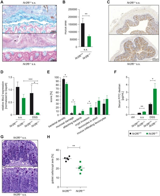
Figure 5.
Nr2f6−/− colons have impaired mucus barrier integrity. (A) Steady-state (s.s.) 10- to 12-week-old wild-type and Nr2f6−/− colonic tissues were fixed with Carnoy’s fixative, sectioned, stained with Alcian blue (blue) and (B) analysed calculating the mucus area using ImageJ (n=8, p=0.0091). (C) Representative images of immunohistochemical staining (brown) with Muc2 antibody in colon sections of steady-state Nr2f6+/+ and Nr2f6−/− mice (n=8). (D) Relative amount of Muc2 mRNA detected in colonic scrapings by quantitative real-time PCR (Nr2f6−/− s.s. versus DSS p=0.0002; wild type versus Nr2f6−/− DSS p=0.021). Data are expressed as relative fold change between Nr2f6+/+ and Nr2f6−/− mice at baseline (n=8–11). (E) Colon sections of 10-week-old s.s. mice were probed with an Alexa Fluor 555-conjugated pan-bacterial EUB338 probe for fluorescence in situ hybridisation (FISH) (n=8) and scored for the indicated features as described in detail in the online supplementary Material and methods section. Data are presented as percent involvement according to the respective genotype (n=8; bacterial density p=0.036, mucosal damage p=0.017, crypt invasion p=0.04). (F) Serum concentrations of fluorescein isothiocyanate (FITC) dextran were measured 4 hours after oral administration to Nr2f6+/+ and Nr2f6−/− mice in the s.s. as well as on day 7 after DSS induction (n=5 10; s.s. p=0.005, DSS p=0.022). (G) Representative images of PAS-stained histological colon samples from healthy 10-week-old Nr2f6+/+ and Nr2f6−/− mice showing significantly decreased goblet cell counts per crypt (p=0.0023) in Nr2f6-deficient mice (H). Data are presented as mean±SEM error bars and are representative of at least two independent experiments. Unpaired Student’s t-test, * p<0.05.

