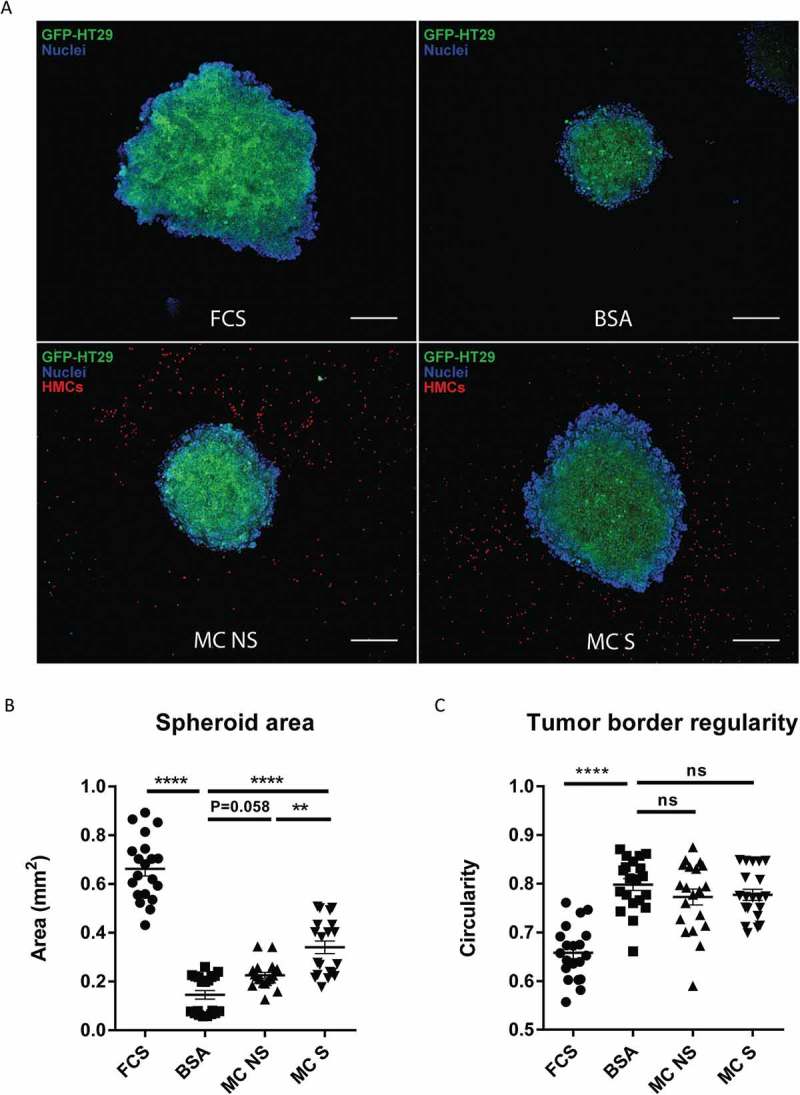
Figure 5.

TLR2-stimulated MC induce stronger growth of colon cancer in a 3D spheroid model.
(a) Representative confocal images of HT29 cancer spheroids cultured in 10% FCS medium, 1% BSA medium or 1% BSA medium in presence of non-stimulated human MC (MC NS) or FSL-1 stimulated human MC (MC S) for 6 days. GFP transfected HT29 cells are shown in green, cell nuclei are shown in blue and CMTPX-labeled MC in red. (Scale bar, 200μm). (b) Spheroid area of HT29 as an indicator of cancer growth. (c) Border regularity of HT29 spheroid as an indicator of cancer invasiveness. Border regularity was calculated in a formula: 4π (spheroid area/spheroid perimeter^2). Data are displayed as mean ± SEM of two independent experiments (n=2) .**P ˂ 0.005; **** P < 0.0001, assessed by one-way ANOVA, Tukey’s multiple comparisons.
