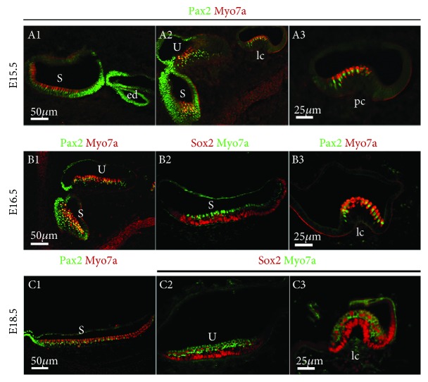
Figure 3.
Comparison of Pax2 and Sox2 gene expression in the vestibule. Immunofluorescence of Pax2, Sox2, and myosin7a in the vestibule from E15.5 to 18.5. S: saccule; U: utricle; ed: endolymphatic duct; pc: posterior crista; lc: lateral crista. Scale bars = 50 μm (A1–A2, B1–B2, and C1–C2); 25 μm (A3, B3, and C3).

