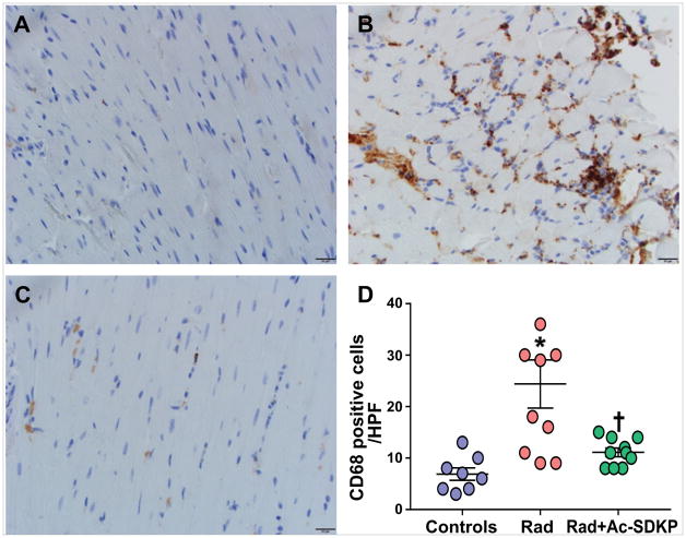
Figure 5. Assessment of myocardial macrophage infiltration.
Panel A–C: Representative images of CD68-stained myocardial macrophages. Panel A: Wild type control; Panel B: Radiation; Panel C: Radiation + Ac-SDKP. Brown stained cells represent macrophages × 400. Panel D: Quantification of myocardial macrophages in control and radiation exposed rats with and without Ac-SDKP therapy. N = 8–10, *, P < 0.001 radiation vs. control, †, P =0.008 radiation vs. radiation + Ac-SDKP.

