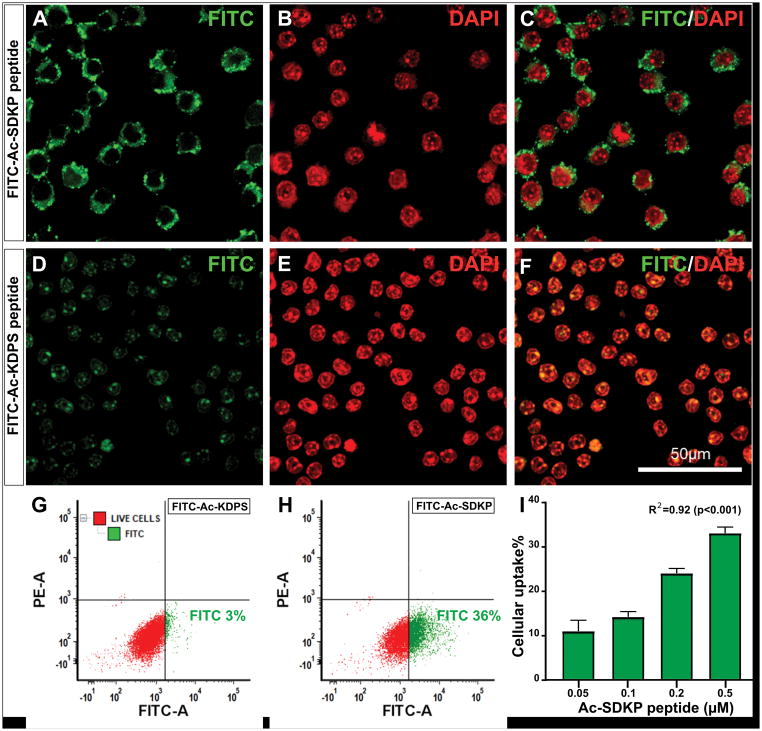
Figure 7. Confocal microscopy images showing the uptake and localization of FITC-Ac-SDKP conjugate into cultured macrophages.
Macrophages were incubated with FITC labelled Ac-SDKP and scrambled peptide for 4 hours and the cells were prepared for immunofluorescence study. A: Representative confocal image showing FITC-labelled peptide (green) localized in to the perinuclear region; B: DAPI stained macrophage nuclei (red), and C: Merged FITC and DAPI images. Panel D–F shows non-specific diffuse and weak cytoplasmic staining of scrambled peptide, FITC-Ac-KDPS (FITC in green and DAPI in red). Panel G–H are representative fluorescence-activated cell sorting (FACS) results corresponding to scrambled peptide and FITC-Ac-SDKP probe, respectively. The gating was set to show FITC-A positive cell population in green and negative viable cells in red. A–H are representative results for macrophages incubated with Ac-SDKP at 0.5μM concentration. Panel I: Bar graph showing percentage of cells with FITC uptake at different concentration of Ac-SDKP with background subtraction of scrambled peptide by FACS analysis. N= 3, R2 = 0.92 (p <0.001). Scale bar:50μm.

