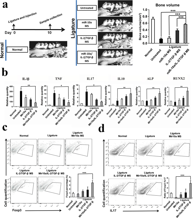
Figure 5. Multi-functionalized PLLA NF-SMS rescued bone resorption in a mouse periodontal disease model.
a) MicroCT results show bone loss between the first and second molars in the periodontitis model and the bone volume changes in various treatment groups; b) The gene expression of gingival tissues was quantified using real-time PCR, showing the effector T cell cytokines IL-1β, TNF, IL17, anti-inflammatory cytokine IL-10, and osteogenic markers ALP and RUNX2; c) The flow cytometry analysis of the isolated gingival tissues show the percentage of Foxp3+ cells (gated on CD4-expressing cells) in different treatment groups. d) The IL17+ cells (gated on CD4-expressing cells) and the ratios of Treg/Th17 cells of the isolated gingival tissues were shown.

