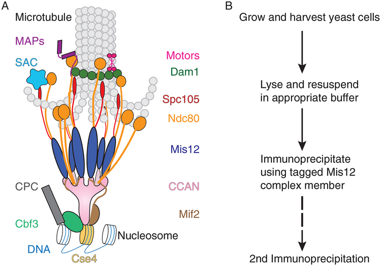
FIG. 1.
Composition and purification of yeast kinetochores. (A) Kinetochores are multiprotein structures that assemble specifically on a specialized nucleosome containing Cse4 (dark yellow). The deposition of Cse4 is facilitated by the Cbf3 complex (aqua) in conjunction with Cbf1 (not shown) and the yeast-specific Cse4 chaperone, Scm3 (not shown). The Cbf3 complex also recruits the chromosomal passenger complex (CPC) (dark gray), which destabilizes kinetochore attachments lacking tension. The Cse4 nucleosome and the adjacent DNA are bound by the larger CCAN (constitutive centromere-associated network) complex (pink) and the Mif2 protein (brown). A conserved interaction between these two facilitates recruitment of the Mis12 complex (dark blue), which brings in the Spc105 (red) and Ndc80 (orange) complexes. Under some conditions, Ndc80 can also be bound by the CCAN, but the bulk of the yeast Ndc80 localizes to the kinetochore via its interaction with the Mis12 complex. The Ndc80 and Spc105 complexes recruit the diffusible wait signal in the event of an unattached kinetochore, the SAC (spindle assembly checkpoint) (teal). Finally, to secure the attachment of the kinetochore to the microtubule, the Ndc80 complex recruits the Dam1 complex (dark green), which oligomerizes to form a ring around the microtubule. The sustained attachment of the kinetochore to the microtubule is mediated by many subcomplexes, including Ndc80, Dam1, and additional microtubule-associated proteins (MAPs) (purple) and molecular motors (hot pink) that transiently localize to the kinetochore as necessary. (B) In order to study the multitude of the kinetochore functions, we have devised a method to purify kinetochores. The workflow involves growing and harvesting cells, lysing them in the appropriate buffer, immunoprecipitating the kinetochores using a tagged component of the Mis12 complex (Dsn1 is preferred), eluting the kinetochores, and, if necessary, performing a second immunoprecipitation. Slight adjustments at every one of these steps of the protocol can alter the composition of the resulting kinetochores.

