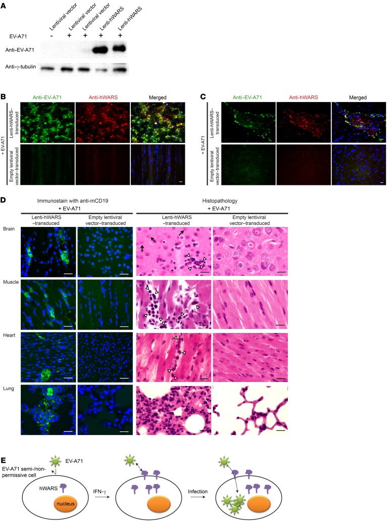
Figure 5. EV-A71 infection in mouse cells overexpressing hWARS.
(A) Western blot analyses of EV-A71 protein in the skeletal muscles of EV-A71–challenged neonatal BALB/c mice pretransduced with empty lentiviral vectors (lanes 2 and 3) or a lentiviral vector expressing hWARS (lanes 4 and 5). Two representative samples were included in each category. A noninfected empty lentiviral vector (lane 1) was included as a control. Representative images of muscle (B) and brain (C) tissues from hWARS-transduced mice. Tissues from EV-A71–infected mice were immunostained with anti–EV-A71 (green) and anti-hWARS antibodies (red), respectively. The tissues from empty lentiviral vector–transduced mice inoculated with EV-A71 served as negative controls. DAPI-stained nuclei (blue) are also shown in the merged images. (D) Histopathology of the EV-A71–inoculated hWARS-transduced mice on day 5 after infection. Interstitial infiltrations of lymphocytes in various organs were detected using anti-CD19 antibodies (green). The nuclei were stained with DAPI (blue). H&E staining showed the inflammatory infiltrates of mononuclear cells (arrowheads) and degenerated neuronal cells (arrows). (E) Schematic representation of an IFN-γ–inducible cell entry model for EV-A71 infection. EV-A71 semi- or nonpermissive cells show low or no surface expression of hWARS in a quiescent state (left). The proinflammatory response leading to the production of IFN-γ can induce the expression and membrane translocation of hWARS. This in turn sensitizes poorly susceptible cells to EV-A71 infection by facilitating hWARS–EV-A71 interaction (middle). As a result, EV-A71 can be internalized into the cells, and infection occurs. The images shown in B–D are representative of 3 independent experiments. Scale bars: 30 μm.

